
The Best, Most Rudimentary Anatomical Diagram
Greetings Scan Squad!
Every day, I see vets doubting their anatomical knowledge!
You all are champs at surgery, knowing where to find certain organs, blood vessels and ligaments. Yet, when it comes to ultrasound, suddenly the gall bladder is located in the caudal abdomen and females have a prostate!
So how do you gain confidence with organ positions?
My best advice: picture where each organ is during an exploratory laparotomy or during a post-mortem or during a dissection lab at university. This is particularly helpful if you perform your ultrasounds in dorsal recumbency, but it will still work for those lateral scanners amongst us!
Secondly, you need to practice.
If you simply cannot find an organ, that is okay! Always write this in your ultrasound report summary, and keep practicing. The easiest way to “find time” to practice, is to block out an extra 15-30 (heck even 10) minutes to the schedule for your spey cases. They will need to be clipped anyway! Can’t get away with this much extra time? Spend five minutes practicing on one organ!
If all else fails, my awesome, rudimentary drawing might help! Whilst scanning, I stand on the right side of the patient (the left side of the image), facing their head.
A final pro tip: if your patient has had a splenectomy in the past, it won’t have a spleen! Even if I’ve been told that there won’t be a spleen, I often start looking for one out of habit, often uttering a “what the hell?”, before I remember! Make sure to review the history and ask your client!
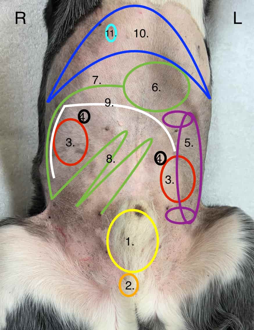
A very rudimentary diagram of organ placement. You know where these organs are, trust yourself!
Picture holding your probe in this general location for each respective organ.
1. Urinary bladder
2. Prostate
3. L and R kidneys
4. L and R adrenal glands
5. Spleen
6. Stomach
7. Duodenum
8. GIT (even more imagination needed here!)
9. Pancreas
10. Liver
11. Gall bladder
Happy scanning!
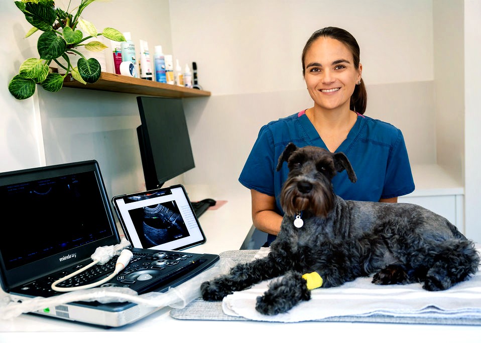
About Belinda
Dr. Belinda Carey is a veterinarian, educator and founder of eVet Academy, with a passion for making veterinary ultrasound practical and accessible. After graduating with first-class honours and earning her MANZCVS in Small Animal Internal Medicine, Belinda spent years providing mobile ultrasound services to general practice clinics, where she recognised a widespread need for practical, step-by-step ultrasound training.
Combining her veterinary expertise with her teaching background, she created eVet Academy to help small animal vets build confidence and improve their scanning skills through clear, clinically relevant education designed for everyday practice.
Browse our other articles
Pathology or Artifact?
Pathology or Artifact? Greetings Scan Squad! One of my instagram posts last week highlighted the importance of testicular scanning. “Hidden” within these images was a very cool ultrasound artifact… …edge shadowing! What is edge shadowing? It is a narrow,...
3 Ultrasound Tips Anyone Can Master
3 Ultrasound Tips Anyone Can Master Greetings Scan Squad! Here are three easy tips that you can do to improve your ultrasound quality, no matter your skill level! Quality imaging starts with the basics. 1.Sedate, sedate, sedate! A relaxed, still patient is key to...
Ultrasound Knobology
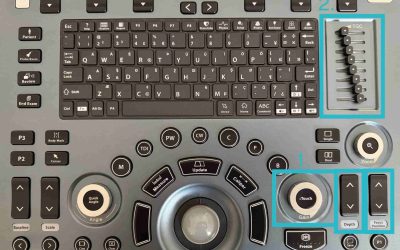
Ultrasound Knobology Greetings Scan Squad! It’s time to review everyone’s favourite (insert intense sarcasm) topic…machine control settings, also known as knobology! Many vets don’t take the time to learn what these dials do because, quite simply, it’s a pretty boring...