
How Important are Ultrasound Reports in Veterinary Ultrasound?
Greetings Scan Squad!
Does your clinic have a standard reporting system for ultrasounds?
A written report, that documents your findings, as well as your interpretation of those findings, is essential. This should be included within your patient’s record. To increase your standard of care, this report should be consistent between the veterinarians in your clinic. It should contain several components:
1. Each organ should be assessed and described based upon organ size, shape, echogenicity and whether it appears normal or abnormal. A description of all abnormal findings (once again, using size, shape and echogenicity) and their locations should be included.
2. Make a note of anything that may have impacted your scan! Have you ever considered documenting factors that may have affected your ability to obtain good image clarity? Did the patient have a large volume ascites or significant peritonitis with fluid or inflammation obscuring your image? Did you look for the medial iliac lymph nodes but couldn’t find them? Chances are, if you can’t find them, they’re normal and you should make a note that you looked and they weren’t seen! Was it a deep-chested dog, making the liver difficult to assess? Were you not able to sedate the patient due to medical concerns?
3. A summary of your findings, complete with your interpretation, differentials and recommendations.
4. A description of any ultrasound-guided procedures that were performed. Did you collect a cystocentesis, FNA thoraco/abdominocentesis?
Don’t forget to save labelled images of each organ system, even if they appear normal! Our ultrasound guide helps identify which images to collect.
You can always create your own template. However, I remember what it was like to be a time-poor clinician, so I thought I would create a template for you, so you can go ahead and take that toilet break, finish your cold coffee or have your 4pm ‘lunch break’! Find our completed template HERE. Even better if you can nab it whilst it’s still on sale!
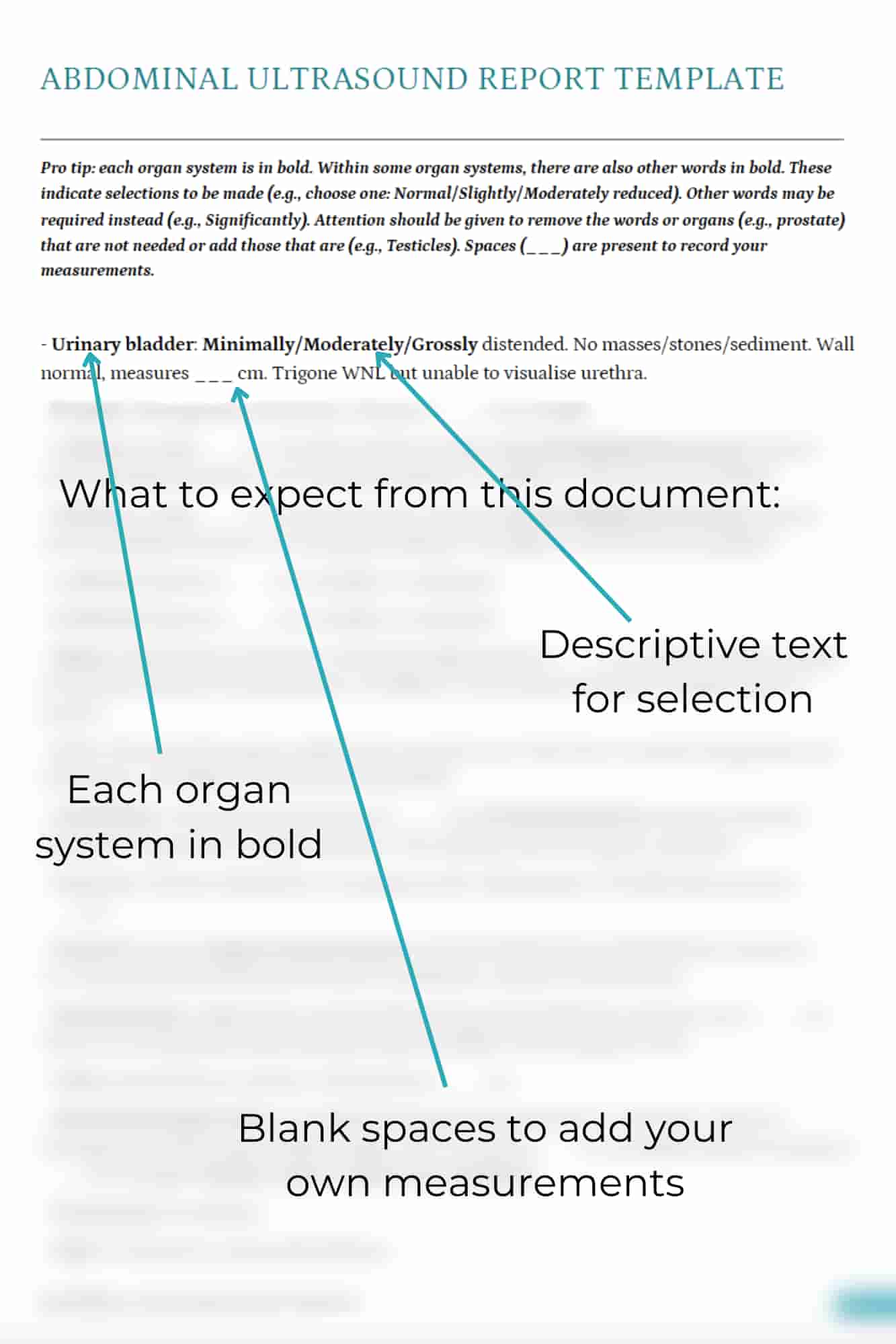
Each Report Template includes a ‘fill-in-the-blank’ ultrasound template and two example ultrasound reports. The completed reports are two of my own actual reports and include the descriptive report, interpretation of the findings and corresponding images.
The below image is a sample of the ultrasound report template – ready to go, saving you tons of time!
About Belinda
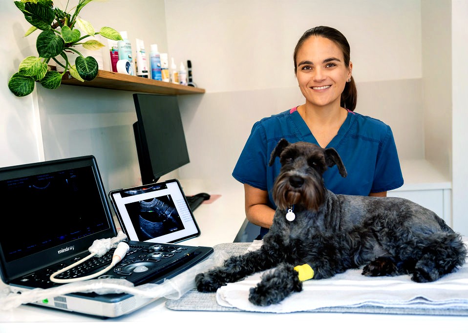
Dr. Belinda Carey is a veterinarian, educator and founder of eVet Academy, with a passion for making veterinary ultrasound practical and accessible. After graduating with first-class honours and earning her MANZCVS in Small Animal Internal Medicine, Belinda spent years providing mobile ultrasound services to general practice clinics, where she recognised a widespread need for practical, step-by-step ultrasound training.
Combining her veterinary expertise with her teaching background, she created eVet Academy to help small animal vets build confidence and improve their scanning skills through clear, clinically relevant education designed for everyday practice.
Browse our other articles
Pathology or Artifact?
Pathology or Artifact? Greetings Scan Squad! One of my instagram posts last week highlighted the importance of testicular scanning. “Hidden” within these images was a very cool ultrasound artifact… …edge shadowing! What is edge shadowing? It is a narrow,...
3 Ultrasound Tips Anyone Can Master
3 Ultrasound Tips Anyone Can Master Greetings Scan Squad! Here are three easy tips that you can do to improve your ultrasound quality, no matter your skill level! Quality imaging starts with the basics. 1.Sedate, sedate, sedate! A relaxed, still patient is key to...
Ultrasound Knobology
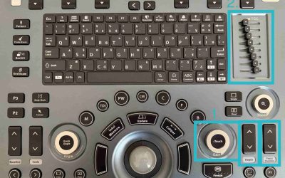
Ultrasound Knobology Greetings Scan Squad! It’s time to review everyone’s favourite (insert intense sarcasm) topic…machine control settings, also known as knobology! Many vets don’t take the time to learn what these dials do because, quite simply, it’s a pretty boring...