
A Cool Kidney
Greetings Scan Squad!
Thanks for hanging around for our second issue of The Probe-Cast!
Who can relate?
You get home from another hard day at work. You’re exhausted. After decompressing, you remember you had the coolest case and want to share the details (and hopefully images!) with your significant other (s). For most of us, I think this typically occurs during dinner time…also known as: the perfect time to pull out the gory stories!
You’re animated as you explain the case details and show your gruesome images. You then look up and see a table of blank or disgusted faces (insert: partners, children, in-laws, pets, whoever will listen!). Your face drops and you sadden when you realise that they have no idea what you’re talking about and/or don’t care and/or have stopped eating.
This is also the way of it in my household, except my ‘gory’ images are always ultrasound images. My husband has absolutely no chance at interpreting an ultrasound image, but he somewhat tries to appear engaged. However, his real talent comes in the form of image imagination – kind of like finding animals in the clouds as children or indicating whether you see a butterfly or a murderer on an inkblot test. When I showed him the image below, he instantly identified a dog, complete with saggy ears, a little black nose and a bright tongue!
Even though I know he has no idea what I’m talking about, I’ll still continue to share my stories and images with him. Not in the hopes that he’ll learn or understand my cases, but for the fun-spin, on an otherwise sad, case!
Keep sharing your stories!
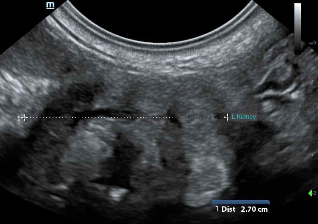
This image actually depicts the left kidney in a 5 MO FE Golden Retriever. Her left kidney measured only 2.7cm in length.
This patient had always been a ‘poor-doer’ since acquired at 8 weeks of age. She had a history of intermittent diarrhoea and anorexia. Although her appetite had recently improved, she had started vomiting and had become lethargic. She had a mild, non-regenerative anaemia. Her biochemistry showed a severe azotaemia, hyperphosphataemia and hypochloraemia. Her right kidney, although larger at 4.9cm in length, also appeared significantly abnormal with a complete loss of renal architecture. A presumed diagnosis of congenital renal dysplasia was made. Her owners declined further investigation.
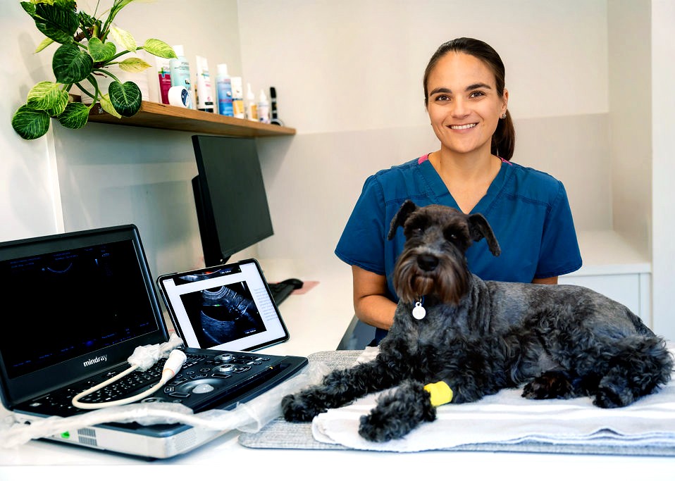
About Belinda
Dr. Belinda Carey is a veterinarian, educator and founder of eVet Academy, with a passion for making veterinary ultrasound practical and accessible. After graduating with first-class honours and earning her MANZCVS in Small Animal Internal Medicine, Belinda spent years providing mobile ultrasound services to general practice clinics, where she recognised a widespread need for practical, step-by-step ultrasound training.
Combining her veterinary expertise with her teaching background, she created eVet Academy to help small animal vets build confidence and improve their scanning skills through clear, clinically relevant education designed for everyday practice.
Browse our other articles
Pathology or Artifact?
Pathology or Artifact? Greetings Scan Squad! One of my instagram posts last week highlighted the importance of testicular scanning. “Hidden” within these images was a very cool ultrasound artifact… …edge shadowing! What is edge shadowing? It is a narrow,...
3 Ultrasound Tips Anyone Can Master
3 Ultrasound Tips Anyone Can Master Greetings Scan Squad! Here are three easy tips that you can do to improve your ultrasound quality, no matter your skill level! Quality imaging starts with the basics. 1.Sedate, sedate, sedate! A relaxed, still patient is key to...
Ultrasound Knobology
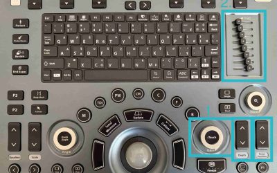
Ultrasound Knobology Greetings Scan Squad! It’s time to review everyone’s favourite (insert intense sarcasm) topic…machine control settings, also known as knobology! Many vets don’t take the time to learn what these dials do because, quite simply, it’s a pretty boring...