
Gastrointestinal Wall Layering = “Bacon”
Greetings Scan Squad!
This week’s topic is also my favourite question to ask final year vet students: what are the four layers of the gastrointestinal tract?
To be fair, most students do very well! However, when asked if they can show me those layers on ultrasound, they’re more stupefied than all of Harry Potter’s enemies combined. This also holds true for many graduated vets, and ironically “which layer is which?” is one of the most common questions I get asked whilst scanning in clinics.
I like to think of the small intestine as the “bacon” of the abdomen. The gastrointestinal wall layering has a similar appearance as the fatty layering in bacon. Whatever helps right?
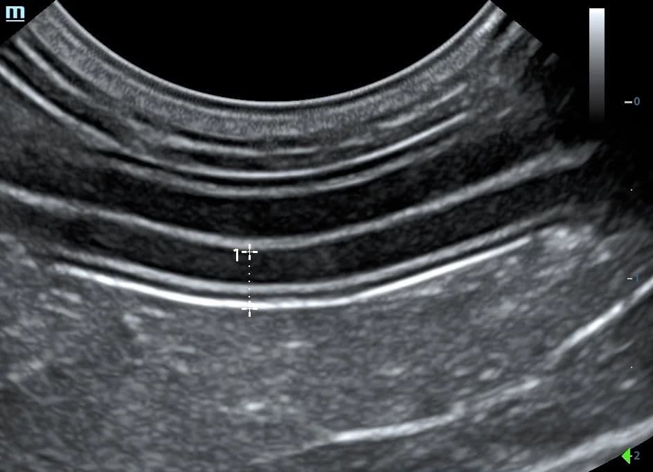
In a ‘normal’ patient, the layers should have a standard, alternating echogenicity. The mucosa will appear hypoechoic (darker) or nearly anechoic (black), the submucosa is hyperechoic (brighter), the muscularis layer is hypoechoic and the serosa is hyperechoic. The lumen will typically be isoechoic (medium grey), but it may be anechoic if liquid contents are present. If there is gas present within the lumen, this will obscure visualisation of the lumen and cause a dirty shadow. The gas will appear as a hyperechoic line along the mucosal surface (arrow in the second image).
Wall height is also very important – make sure you sign up to the mailing list on our website to receive your FREE quick reference measurement guide, which includes the normal wall height for the stomach, duodenum, jejunum, ileum and colon, in both dogs and cats. Yes, of course they are different! Pro tip: always measure wall thickness where you can see all four layers clearly.
Once you start appreciating the normal appearance of the gastrointestinal wall, you can then start identifying abnormalities, such as muscularis layer thickening or the presence of gas within the mucosa itself. A topic for another day!
Happy scanning!
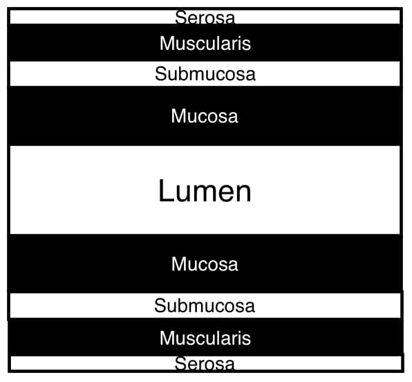
The black and white used in this diagram roughly corresponds to each layer’s appearance on ultrasound.
About Belinda
Dr. Belinda Carey is a veterinarian, educator and founder of eVet Academy, with a passion for making veterinary ultrasound practical and accessible. After graduating with first-class honours and earning her MANZCVS in Small Animal Internal Medicine, Belinda spent years providing mobile ultrasound services to general practice clinics, where she recognised a widespread need for practical, step-by-step ultrasound training.
Combining her veterinary expertise with her teaching background, she created eVet Academy to help small animal vets build confidence and improve their scanning skills through clear, clinically relevant education designed for everyday practice.
Browse our other articles
Pathology or Artifact?
Pathology or Artifact? Greetings Scan Squad! One of my instagram posts last week highlighted the importance of testicular scanning. “Hidden” within these images was a very cool ultrasound artifact… …edge shadowing! What is edge shadowing? It is a narrow,...
3 Ultrasound Tips Anyone Can Master
3 Ultrasound Tips Anyone Can Master Greetings Scan Squad! Here are three easy tips that you can do to improve your ultrasound quality, no matter your skill level! Quality imaging starts with the basics. 1.Sedate, sedate, sedate! A relaxed, still patient is key to...
Ultrasound Knobology
Ultrasound Knobology Greetings Scan Squad! It’s time to review everyone’s favourite (insert intense sarcasm) topic…machine control settings, also known as knobology! Many vets don’t take the time to learn what these dials do because, quite simply, it’s a pretty boring...