
Veterinary Ultrasound Renal Anatomy
Greetings Scan Squad!
In last week’s edition of The Probe-Cast, we reviewed a case of renal dysplasia in a 5 month old Golden Retriever. The architecture of this kidney was very abnormal with no normal tissue visible (see below). Subsequently, a request to review the ultrasonographic appearance of normal renal architecture was made by one of our readers. So let’s get into it!
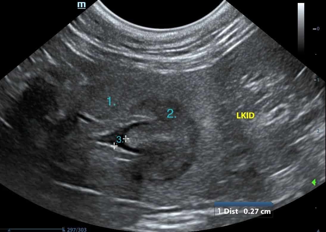
Below, there are two images of an ultrasonographically normal left kidney. The top image shows the kidney in longitudinal view and the bottom image shows the kidney in transverse view. For visualisation purposes, it can help to compare the kidney to a cut tomato!
The kidneys are surrounded by a capsule (the skin of the tomato). On ultrasound, this is usually visualised as a hyperechoic (bright) rim surrounding the renal tissue. The arrow in the first image is pointing to the capsule.
Adjacent to the capsule is the renal cortex (the flesh of the tomato). The cortex contains the glomeruli and convoluted tubules. It should be uniformly echogenic (the same ‘colour’ throughout) and hyperechoic to the medulla.
Next, the inner portion of the kidney corresponds to the medulla (the seeds of the tomato). This area contains the renal pyramids and should be hypoechoic (dark) to the renal cortex. It is the margin between the cortex and medulla where you assess the corticomedullary junction (CMJ). A reduction in CMJ may correspond to renal disease.
Finally, the renal pelvis (the core of the tomato). This is the central collecting system, where urine collects before entering the ureters for transport to the urinary bladder. As a result, this area should be anechoic (without echo = black) in a normal patient because it contains fluid. Minimal dilation of the pelvis should be present and a normal ureter will not usually be visible. We delve into how to measure the renal pelvis as well as normal measurements in our guide book.
If you ever have any newsletter or instagram topic requests, let us know!
Don’t forget to follow us on Instagram.
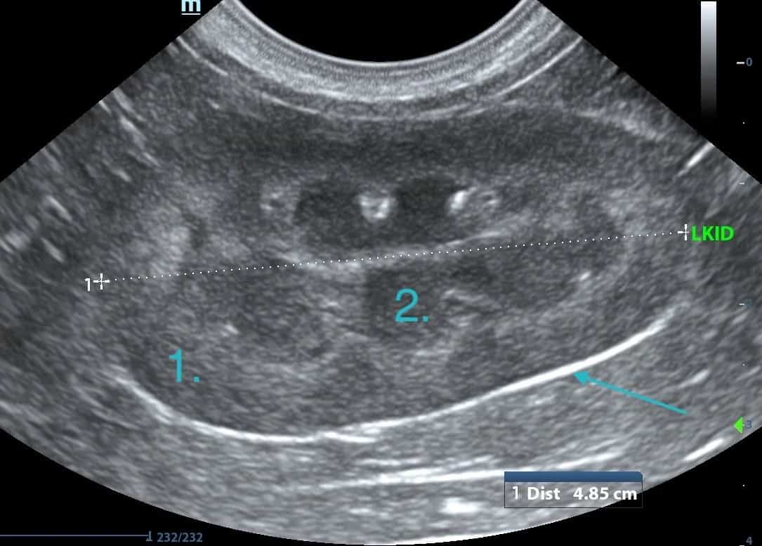
This image depicts the left kidney in longitudinal view and the below image shows the kidney in transverse view.
The arrow is indicating the renal capsule.
1. Cortex
2. Medulla
3. Renal pelvis.
About Belinda
Dr. Belinda Carey is a veterinarian, educator and founder of eVet Academy, with a passion for making veterinary ultrasound practical and accessible. After graduating with first-class honours and earning her MANZCVS in Small Animal Internal Medicine, Belinda spent years providing mobile ultrasound services to general practice clinics, where she recognised a widespread need for practical, step-by-step ultrasound training.
Combining her veterinary expertise with her teaching background, she created eVet Academy to help small animal vets build confidence and improve their scanning skills through clear, clinically relevant education designed for everyday practice.
Browse our other articles
Pathology or Artifact?
Pathology or Artifact? Greetings Scan Squad! One of my instagram posts last week highlighted the importance of testicular scanning. “Hidden” within these images was a very cool ultrasound artifact… …edge shadowing! What is edge shadowing? It is a narrow,...

3 Ultrasound Tips Anyone Can Master
3 Ultrasound Tips Anyone Can Master Greetings Scan Squad! Here are three easy tips that you can do to improve your ultrasound quality, no matter your skill level! Quality imaging starts with the basics. 1.Sedate, sedate, sedate! A relaxed, still patient is key to...

Ultrasound Knobology
Ultrasound Knobology Greetings Scan Squad! It’s time to review everyone’s favourite (insert intense sarcasm) topic…machine control settings, also known as knobology! Many vets don’t take the time to learn what these dials do because, quite simply, it’s a pretty boring...